Medical imaging plays a crucial role in diagnosing, treating, and monitoring health conditions. Whether you’ve experienced an injury, are managing a chronic illness, or are recovering after surgery, imaging gives physicians a clear, noninvasive view inside the body to support more precise care.
At Gaylord Specialty Healthcare, imaging is integrated into the rehabilitation process. Our specialists work closely with referring physicians and radiology teams to ensure every image contributes to a clearer understanding of your condition and a more personalized treatment plan.
This guide explains the most common types of medical imaging, when each is used, and what patients should know before their scan.
What Is Medical Imaging?
Medical imaging allows clinicians to view structures and processes inside the body using noninvasive technology. Each imaging method, such as MRI, CT, ultrasound, or X-ray, provides different kinds of information, depending on what part of the body is being studied.
When deciding which test is best for you, your care team considers several factors:
- What needs to be seen: Some scans are best for visualizing bones and joints, while others are designed to capture soft tissues like muscles, organs, or the brain.
- How much detail is needed: Higher-resolution scans can reveal small structures and subtle changes that help your care team make a more accurate diagnosis.
- Exposure and safety: Some imaging tests use radiation, while others such as MRI and ultrasound do not. Your team always selects the safest option for your needs.
- Use of contrast dye: Certain scans use contrast agents, such as a special dye, to make specific tissues, blood vessels, or organs easier to see.
Each type of imaging has its own strengths and limitations. Understanding how they differ helps patients and clinicians choose the most effective test for each stage of care.
Major Types of Medical Imaging
X-Ray (Radiography)
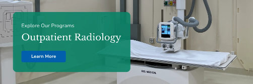
X-rays are one of the most common and widely used imaging tools in medicine. They use a small amount of radiation to create two-dimensional images that show structures inside the body, especially bones.
Key things to know:
- Strengths: Quick, inexpensive, and excellent for identifying fractures, joint alignment, and chest conditions.
- Common Uses: Diagnosing bone fractures, evaluating lung and chest issues, and checking spinal alignment.
- Safety: Radiation exposure is very low, but unnecessary repeat scans are avoided whenever possible.
CT Scan (Computed Tomography)
A CT scan combines a series of X-ray images taken from different angles to create detailed, cross-sectional pictures of the body. It provides more information than a traditional X-ray and is especially valuable in emergency or trauma settings.
Key things to know:
- Strengths: Produces high-resolution images that help detect bone injuries, bleeding, or internal organ conditions.
- Common Uses: Assessing trauma, evaluating bone or joint injuries, and diagnosing lung or abdominal issues.
- Safety: CT scans use more radiation than standard X-rays. Some scans require contrast dye to highlight certain areas, and drinking fluids afterward helps flush it from your body.
MRI (Magnetic Resonance Imaging)
An MRI uses powerful magnets and radio waves to create detailed images of soft tissues inside the body. It’s especially helpful for evaluating the brain, spinal cord, muscles, ligaments, and joints. Because MRI doesn’t use radiation, it’s often preferred for repeat or long-term imaging.
Key things to know:
- Strengths: Provides exceptional detail for soft tissues and neurological structures.
- Common Uses: Helps diagnose spine and joint conditions, ligament or tendon injuries, and brain or nerve disorders.
- Safety: MRI is safe for most patients. You’ll be asked to remove any metal items and talk with your care team about implants before your scan.
Ultrasound (Sonography)
Ultrasound uses high-frequency sound waves to create real-time images of soft tissues and organs. It’s a versatile tool that allows providers to see movement, blood flow, and internal structures without radiation.
Key things to know:
- Strengths: Safe, portable, and radiation-free. It provides real-time images and can guide certain medical procedures.
- Common Uses: Evaluating blood flow, muscle and tendon health, organ function, and guiding injections or treatments.
- Safety: Ultrasound is one of the safest imaging methods and can be used for patients of all ages.
Nuclear Medicine and PET/SPECT Scans
Nuclear medicine uses small amounts of radioactive materials, called tracers, to show how organs and tissues are functioning. PET and SPECT scans are advanced forms of nuclear imaging that measure metabolism and activity in the body.
Key things to know:
- Strengths: Shows how organs and tissues are working, not just how they look.
- Common Uses: Detecting cancer, evaluating heart or bone conditions, and monitoring organ function.
- Safety: Radiation exposure is minimal, but your care team may recommend avoiding close contact with others for a short time after your test.
Hybrid and Interventional Imaging
Some imaging technologies combine multiple methods to give doctors a more complete view of the body. For example, PET-CT and PET-MRI scans merge functional and structural data to provide a more comprehensive picture of the body. Other forms, such as fluoroscopy, provide real-time imaging that allows clinicians to guide treatments or monitor movement as it happens.
Key things to know:
- Purpose: Combines imaging methods to capture more information and improve accuracy in diagnosis and treatment.
- Benefits: Provides greater detail, supports minimally invasive procedures, and helps clinicians make safer, more informed decisions.
How to Choose the Right Imaging Modality
Choosing the right imaging test depends on your symptoms, medical history, and safety factors such as pregnancy, implants, or claustrophobia. Your doctor will also consider image quality, test availability, and insurance coverage to ensure the scan provides the clearest results in the safest and most efficient manner.
Ultimately, the goal is to balance accuracy, safety, comfort, and access while choosing the imaging approach that best supports your diagnosis and treatment plan.
What Patients Should Know Before an Imaging Exam
Preparing properly helps ensure clear results and a comfortable experience. You may need to remove metal objects, fast beforehand, or stay hydrated, depending on the test. If contrast dye is used, your care team will review any allergies or conditions that could affect safety.Imaging exams are carefully designed with patient safety in mind. Radiation exposure is kept as low as possible, and contrast agents are used only when necessary. Be sure to tell your care team if you’re pregnant, have metal implants, or have had a reaction to contrast dye in the past.
At Gaylord Specialty Healthcare, imaging is part of a complete approach to recovery. Our specialists use advanced technology and a collaborative process to make each scan as safe and effective as possible. Learn more about our imaging and rehabilitation services Learn more about our imaging and rehabilitation services to see how we help patients move forward with confidence.
This content is for educational purposes only and is meant to provide general information. It is not a substitute for professional medical advice, diagnosis, or treatment. Always consult your healthcare provider with any questions or concerns about your health. In case of a medical emergency, contact your doctor or call 911 right away.